In today’s medical imaging field, clarity is everything. Advanced MRI, CT, X-ray, and ultrasound systems now offer highly refined greyscale gradients that let radiologists see even the smallest differences in tissue structure. These subtle shifts from deep blacks to soft mid-tones and bright whites help identify early abnormalities, reduce uncertainty, and improve diagnostic accuracy.
Understanding Greyscale Imaging
Greyscale imaging works by converting internal body signals into simple shades of black, white, and grey. Each shade shows how much signal a tissue absorbs or emits, giving radiologists the contrast they need to spot what’s normal and what isn’t.
What the shades tell us:
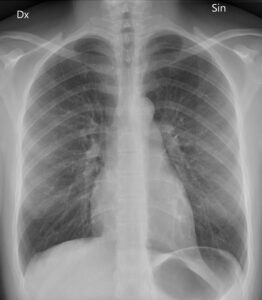
- Bright tones (near white): Dense structures like bone or calcifications.
- Mid-grey tones: Soft tissues such as organs, muscles, and fat.
- Dark tones (near black): Air or fluid-filled areas, like lungs or cysts.
The real strength of greyscale imaging is its precision. Modern scanners can capture extremely fine differences in tone—details too subtle for the human eye alone. This level of clarity helps clinicians detect issues earlier, interpret images with more confidence, and deliver better patient care.
Greyscale Diagnostic Significance
This precision is what gives greyscale imaging its strong diagnostic significance. By revealing subtle variations in tissue density and structure, greyscale scans provide radiologists with the clarity needed to identify abnormalities at an early stage and make more confident clinical decisions.
Key diagnostic advantages include:
- Precise tissue differentiation – for identifying soft tissue changes, fluid, inflammation, or masses.
- High contrast and clarity – which make structural edges, textures, and abnormalities easier to see.
- Early detection capability – thanks to fine tonal gradients that reveal subtle signs of disease.
- Reliable visualization – of bones, organs, vessels, and fluid-filled areas.
- Greater diagnostic confidence – by reducing ambiguity in interpretation.
- Consistency across modalities – such as MRI, CT, X-ray, and ultrasound.
Applications Of Greyscale Imaging in Medical Diagnosis
Because of its exceptional precision, greyscale imaging is vital across a wide range of clinical applications. By converting complex internal structures into clean, high-contrast visuals it allows radiologists to assess bones, soft tissues, organs, and vessels with speed and accuracy. This clarity supports earlier detection, more confident interpretation, and improved diagnostic outcomes. Below is an overview of the key areas where greyscale imaging plays a crucial role in medical diagnosis.
Key Applications of Greyscale Imaging
| Sr | Application Area | How Greyscale Imaging Helps | Common Modalities |
|---|---|---|---|
| 1 | Bone / Trauma Assessment | Reveals fractures, deformities, bone lesions, bleeding, and internal injuries | X-ray, CT |
| 2 | Soft Tissue Evaluation | Differentiates organs, muscles, and fat to detect abnormalities | MRI, CT, Ultrasound |
| 3 | Tumor & Lesion Detection | Highlights subtle tonal changes that indicate masses or cysts | MRI, CT, Ultrasound |
| 4 | Cardiac Imaging | Shows heart structure, chamber size, and tissue changes | MRI, Echocardiography |
| 5 | Neurological Imaging | Visualizes brain anatomy, swelling, bleeds, and density variations | MRI, CT |
| 6 | Lung & Chest Imaging | Distinguishes air, fluid, and tissue to detect infections or masses | X-ray, CT |
| 7 | Abdominal & Pelvic Imaging | Identifies organ enlargement, stones, fluid buildup, and soft tissue issues | CT, MRI, Ultrasound |
| 8 | Vascular Imaging | Displays vessel walls, blockages, clots, and flow patterns | Ultrasound (Doppler), CT Angiography |
| 9 | Spine Evaluation | Shows disc problems, fractures, and spinal canal narrowing | MRI, CT |
| 10 | Prenatal & Obstetric Imaging | Supports fetal growth monitoring and anatomy assessment | Ultrasound |
| 11 | Inflammation & Infection Detection | Shows swelling, abscesses, and structural changes | MRI, CT, Ultrasound |
| 12 | Guided Procedures | Provides real-time visuals for biopsies, drainages, and injections | Ultrasound, CT |
How to Get the Best Results from Greyscale Imaging?
Achieving high-quality greyscale images requires more than just advanced equipment depends on proper image processing and consistent calibration. To achieve optimal diagnostic quality, greyscale images are often enhanced through techniques like windowing (adjusting brightness and contrast), filtering (reducing grain while preserving detail), and image sharpening, all of which help improve diagnostic clarity. DICOM Standardization across imaging systems ensures that greyscale values remain consistent, regardless of modality or workstation. With advancements in AI and machine learning, greyscale image analysis is becoming more accurate, enabling automatic detection of abnormalities and improving diagnostic confidence.
How to Print Those Results Accurately Using Halftones?
Although greyscale and halftones serve different functions, they are closely connected through how tonal information is represented and reproduced. Greyscale carries true, continuous tonal data used directly for diagnosis. Halftones, on the other hand, are used to simulate those greyscale tones on physical media.
Greyscale delivers the diagnostic precision radiologists rely on, while halftones make that precision reproducible on physical media. Together, they ensure accurate, reliable communication of medical information—whether viewed digitally or in print.
Challenges in Greyscale Imaging
Despite its diagnostic value, greyscale imaging faces several practical and technical limitations:
1. Tonal Accuracy & Consistency
Reliable diagnosis depends on precise greyscale reproduction. Achieving deep blacks (high D-max) and stable mid-tones can be difficult, especially with inkjet systems that may not match traditional film quality. Color-based scans also face metamerism, where tones shift under different lighting, causing inconsistencies during review.
2. Print Durability
Medical prints must remain stable for long-term records. Dye-based inks can fade with light, humidity, or handling. Pigment inks last longer but may offer a smaller color range or face jetting challenges. Fresh prints can smudge before drying, slowing down clinical workflows.
3. Media Compatibility
Medical imaging requires printing on specially coated transparent films. Inks must adhere properly, maintain high optical density, and avoid bleeding. Creating inks that meet these technical and safety requirements while preserving diagnostic clarity is required to ensure the sanctity of medical imaging.
Overcoming Key Challenges With High-Resolution Medical Image Printing
Modern medical inkjet printing systems are engineered to address the core challenges of greyscale imaging—accuracy, durability, media compatibility, and standardization.
These advanced printers produce high-resolution diagnostic images on specialized medical-grade transparent films, ensuring deep blacks, smooth tonal transitions, and stable mid-tones. This helps radiologists detect subtle fractures, soft-tissue variations, and early abnormalities with greater confidence.
To overcome common limitations:
- Enhanced tonal accuracy ensures consistent grayscale reproduction and minimizes metamerism, allowing images to appear reliable under different lighting conditions.
- High fade-resistant dye-based & pigment inks that improve long-term durability of prints, preventing degradation in storage or clinical environments.
- Optimized ink formulations for sharpness & no inter colour bleeding, delivering high optical density without smudging or bleeding, crucial for fast-paced workflows.
- Full DICOM and regulatory compliance ensures standardized output across systems, making prints dependable for cross-hospital or multi-clinic review.
Conclusion
Modern medical imaging systems, paired with high-quality inkjet printers, offer a cost-effective and reliable alternative to traditional film printing, making diagnostic-grade imaging accessible for clinics, diagnostic centers, and smaller healthcare facilities.
Building on this advancement, Splashjet’s specialized Medical Imaging Inks facilitate consistent grayscale accuracy, deep contrast, and dependable print quality on medical films. With Splashjet, imaging centers and radiology labs can deliver clear, high-precision prints that support confident diagnosis every time.